Showing posts with label Diseases & Conditions. Show all posts

Sunday, June 15, 2014

Ascites

 Definition 

Ascites with umbilical hernia.
Ascites is the accumulation of excess fluid in the peritoneal cavity, most commonly caused by liver cirrhosis.


Physical finding and Clinical presentation

  • Important information to elicit within history
    • Viral hepatitis
    • Alcoholism
    • Increasing abdominal girth, umbilical hernia
    • Increasing lower extremity edema
    • Intravenous drug use
    • Sexual history (i.e., men who have sex with men)
    • History of transfusions
  • Important physical examination findings
    • Bulging flanks
    • Flank dullness to percussion
    • Fluid wave on abdominal examination
    • Lower extremity edema
    • Shifting dullness “succusion splash” on abdominal examination
    • Physical signs associated with liver cirrhosis: spider angiomas, jaundice, loss of
      body hair, Dupuytren’s contracture, muscle wasting, bruising, palmar erythema, gynecomastia, testicular atrophy, hemorrhoids, caput meduse
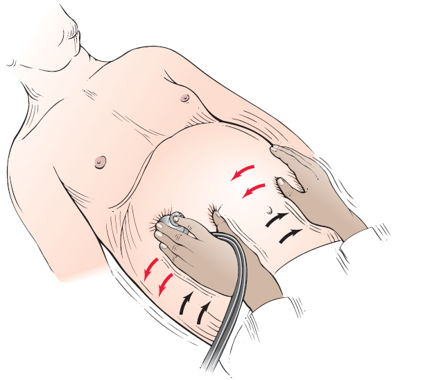
"Succusion splash” technique for assessing distention of abdominal
viscera
.

Cause

Pathophysiology of ascites: increased hepatic resistance to portal flow leads to portal hypertension. The splanchnic vessels respond by increased secretion of nitric oxide causing splanchnic artery vasodilation. Early in the disease increased plasma volume and increased cardiac output compensate for this vasodilation. However, as disease progresses the effective arterial blood volume decreases causing sodium and fl uid retention through activation of the renin-angiotensin system. The change in capillary pressure causes increased permeability and retention of fluid in the abdomen.


Differential Diagnosis

  • Chronic parenchymal liver disease, leading to portal hypertension
  • Peritoneal carcinomatosis
  • Congestive heart failure
  • Peritoneal tuberculosis
  • Nephrotic syndrome
  • Pancreatitis
Laboratory Tests
  • Initial evaluation should always include:
    • Diagnostic paracentesis. Laboratory tests on this fluid should include a CBC with differential, albumin, total protein, culture and a Gram stain. Optional tests on paracentesis fluid (depending on patient’s history) include amylase, LDH, acid-fast bacilli and glucose levels
    • AST, ALT, total and direct bilirubin, albumin, alkaline phosphatase, GGTP
    • CBC, coagulation studies
    • Electrolytes, BUN, creatinine
  • A serum to ascites albumin gradient (SAAG) should be calculated in all patients. If the SAAG is greater than 1.1, the cause of ascites can be attributed to portal hypertension. If SAAG is less than 1.1, a nonportal hypertension etiology of ascites must be sought.
Imaging studies
  • Endoscopy of the upper GI tract to evaluate for esophageal varices if ascites is secondary to portal hypertension.
  • Abdominal ultrasound is the most sensitive measure for detecting ascitic fl uid; a CT scan is a viable alternative.
  • Liver biopsy in select patients (i.e., those with portal hypertension of uncertain etiology).
Treatment
  • Sodium-restricted diet (maximum 60-90 milliequivalents per day).
  • Fluid restriction to 1 liter per day in patients with hyponatremia.
  • Patients with moderate-volume ascites causing only moderate discomfort may be treated on an outpatient basis with the following diuretic regimen: spironolactone 50-200 mg daily or amiloride 5-10 mg daily. Add furosemide 20-40 mg per day in the fi rst several days of treatment, monitoring renal functions carefully for signs of prerenal azotemia (in patients without edema goal weight loss is 300-500 grams/ day, in patients with edema 800-1000 grams/day).
  • Patients with large-volume ascites causing marked discomfort or decrease in activities of daily living may also be treated as outpatients if there are no complications. There are two options for treatment in these patients: (1) largevolume paracentesis or (2) diuretic therapy until loss of fluid is noted (maximum spironolactone 400 mg daily and Lasix 160 mg daily). There is generally no difference in longterm mortality; however paracentesis is faster, more effective,
    and associated with fewer adverse effects.
  • Five percent to 10% of patients with large-volume ascites will be refractory to high-dose diuretic treatment. Treatment strategies include repeated large-volume paracentesis with infusion of albumin every 2-4 weeks or placement of a transjugular intrahepatic portosystemic shunt (TIPS).
Read More

Saturday, June 14, 2014

Appendicitis


Definition

Appendicitis is the acute inflammation of the appendix.

Physical finding and Clinical presentation
  • Abdominal pain: initially the pain may be epigastric or periumbilical in nearly 50% of patients; it subsequently localizes to the RLQ within 12 to 18 hours. Pain can be found in the back or right fl ank if appendix is retrocecal or in other abdominal locations if there is malrotation of the appendix.
  • Pain with right thigh extension (psoas sign), low-grade fever: temperature may be 38° C if there is appendiceal perforation.
  • Pain with internal rotation of the fl exed right thigh (obturator sign) is present.
  • RLQ pain on palpation of the LLQ (Rovsing’s sign): physical examination may reveal right-sided tenderness in patients with pelvic appendix.
  • Point of maximum tenderness is in the RLQ (McBurney’s point).
  • Nausea, vomiting, tachycardia, cutaneous hyperesthesias at the level of T12 can be present.
Cause

Obstruction of the appendiceal lumen with subsequent vascular congestion, inflammation, and edema; common causes of obstruction are:
  • Fecaliths: 30% to 35% of cases (most common in adults)
  • Foreign body: 4% (fruit seeds, pinworms, tapeworms, roundworms, calculi)
  • Inflammation: 50% to 60% of cases (submucosal lymphoid hyperplasia [most common etiology in children, teens])
  • Neoplasms: 1% (carcinoids, metastatic disease, carcinoma)
Differential Diagnosis
  • Intestinal: regional cecal enteritis, incarcerated hernia, cecal diverticulitis, intestinal
    obstruction, perforated ulcer, perforated cecum, Meckel’s diverticulitis.
  • Reproductive: ectopic pregnancy, ovarian cyst, torsion of ovarian cyst, salpingitis, tubo-ovarian abscess, Mittelschmerz endometriosis, seminal vesiculitis.
  • Renal: renal and ureteral calculi, neoplasms, pyelonephritis
  • Vascular: leaking aortic aneurysm
  • Psoas abscess
  • Trauma
  • Cholecystitis
  • Mesenteric adenitis
Laboratory Tests
  • CBC with differential reveals leukocytosis with a left shift in 90% of patients with appendicitis. Total WBC count is generally lower than 20,000/mm3. Higher counts may be indicative
    of perforation. Less than 4% have a normal WBC and differential. A low Hgb and Hct in an older patient should raise suspicion for GI tract carcinoma.
  • Microscopic hematuria and pyuria may occur in 20% of patients.
Imaging Studies
  • CT of the right lower quadrant of the abdomen has a sensitivity of 90% and an accuracy 94% for acute appendicitis. A distended appendix, periappendiceal inflammation, and a thickened appendiceal wall are indicative of appendicitis.
  • Ultrasonography has a sensitivity of 75% to 90% for the diagnosis of acute appendicitis, although it is highly operator dependent and diffi cult in patients with large body habitus. Ultrasound is useful, especially in younger women when diagnosis is unclear. Normal ultrasonographic findings should not deter surgery if the history and physical examination are indicative of appendicitis.
Treatment
  • Urgent appendectomy (laparoscopic or open), correction of fluid and electrolyte imbalance with vigorous IV hydration and electrolyte replacement.
  • IV antibiotic prophylaxis to cover gram-negative bacilli and anerobes.
Read More

Epiglottitis

 
Acutely inflamed epiglottis associated with Haemophilus influenza type B


Definition

Epiglottitis is a rapidly progressive cellulitis of the epiglottis and adjacent soft tissue structures with the potential to cause abrupt airway obstruction.

Physical finding and Clinical presentation
  • Irritability, fever, dysphonia, dysphagia
  • Respiratory distress, with child tending to lean up and forward
  • Often, drooling or oral secretions
  • Often, presence of tachycardia and tachypnea
  • On visualization, edematous and cherry red epiglottis
  • Often, no classic barking cough as seen in croup
  • Possibly fulminant course (especially in children), leading to complete airway obstruction.
Cause
  • In children, Haemophilus infl uenzae type B is usual.
  • In adults, H. infl uenzae can be isolated from blood, epiglottis (about 26% of cases), or both.
  • Pneumococci, streptococci, and staphylococci are also implicated.
  • Role of viruses in epiglottitis unclear.
Differential Diagnosis
  • Croup
  • Angioedema
  • Peritonsillar abscess
  • Retropharyngeal abscess
  • Diphtheria
  • Foreign body aspiration
  • Lingual tonsillitis
Work Up
  • Cultures of blood and urine
  • Lateral neck radiograph to show an enlarged epiglottis, ballooning of the hypopharynx, and normal subglottic structures
    • Radiographs are of only moderate sensitivity and specificity and take time to perform.
    • Visualization of the epiglottitis may be safer in adults than in children. In children, visualization and intubation are best done in the most controlled environment.
  • Cultures of the epiglottitis
Laboratory Tests
  • CBC: may reveal a leukocytosis, with a shift to the left
  • Chest x-ray examination: may reveal evidence of pneumonia in almost 25% of cases
  • Cultures of blood, urine, and epiglottis, as noted
Treatment
  • Maintenance of adequate airway is critical.
  • Early placement of an endotracheal or nasotracheal tube in a child is advised.
  • Closely follow adult patient and defer intubation, provided the airway reveals no signs of obstruction.
  • H. influenzae in children may be less common because of the availability of the Hib vaccine.
  • Use antibiotics such as ceftriaxone, cefotaxime.
Read More

Pharyngitis and Tonsillitis

Definition
Pharyngitis or tonsillitis is inflammation of the pharynx or tonsils.
Physical finding and Clinical presentation
Adenoviral Pharyngitis
  • Pharynx
    1. May appear normal to severely erythematous
    2. Tonsillar hypertrophy and exudates commonly seen but do not indicate cause.
  • Viral infection
    1.  Rhinorrhea
    2. Conjunctivitis
    3. Cough
  • Bacterial infection, especially group A Streptococcus
    1. High fever
    2. Systemic signs of infection
  • Herpes simplex or enterovirus infection: vesicles
  • Streptococcal infection
    • Rare complications
      • Scarlet fever
      • Rheumatic fever
      • Acute glomerulonephritis
    • Extension of infection: tonsillar, parapharyngeal, or retropharyngeal abscess presenting with severe pain, high fever, trismus.
Cause
Viruses
  • Respiratory syncytial virus
  • Influenza types A and B
  • Epstein-Barr virus
  • Adenovirus
  • Herpes simplex
Bacteria
  • Streptococcus
  • Neisseria gonorrhoeae
  • Arcanobacterium haemolyticum
Other organisms
  • Mycoplasma pneumoniae
  • Chlamydia pneumoniae
Differential Diagnosis
  • Sore throat associated with granulocytopenia, thyroiditis
  • Tonsillar hypertrophy associated with lymphoma.
Workup
  • Throat swab for culture to exclude S. pyogenes, N. gonorrhoeae (requires specifi c transport medium)
  • Rapid streptococcal antigen test (culture should be performed if rapid test negative and diagnosis suspected)
  • Monospot
Laboratory Tests
  • CBC with differential
    • May help support diagnosis of bacterial infection
    • Streptococcal infection suggested by leukocytosis of more
      than 15,000/mm3
  • Viral cultures, serologic studies rarely needed
Imaging Study
  • Seldom indicate
Treatment
  • If streptococcal infection proved or suspected:
    • Penicillin
    • Erythromycin if penicillin allergic
  • If gonococcal infection proved or suspected: ceftriaxone
  • Tonsillopharyngitis is generally managed in an outpatient setting with follow-up arranged in a week or two. Admission to the hospital is indicated for local suppurative complications (peritonsillar abscess, lateral pharyngeal or posterior pharyngeal abscess, impending airway closure, or inability to swallow food, medications, or water).
Pharyngitis associated with GAS (Group A strep) infection


    Read More

    Friday, June 13, 2014

    Bell's Palsy

    Definition
    Fig. 1: Right Facial Palsy


    Bell’s palsy is an idiopathic, isolated, usually unilateral facial weakness in the distribution of the seventh cranial nerve; less than 1% are bilateral.

    Physical finding and Clinical presentation
    • Unilateral paralysis of the upper and lower facial muscles (asymmetrical eye closure, brow, and smile, Fig.1).
    • Ipsilateral loss of taste
    • Ipsilateral ear pain, usually 2 to 3 days before presentation
    • Increased or decreased unilateral eye tearing
    • Hyperacusis
    • Subjective ipsilateral facial numbness
    • In about 8% of cases, other cranial neuropathies may occur.
    Cause
    • Most cases are idiopathic, although the cause is often viral (herpes simplex).
    • Herpes zoster can cause Bell’s palsy in association with herpetic blisters affecting the outer ear canal or the area behind the ear (Ramsay-Hunt syndrome).
    • Bell’s palsy can also be one of the manifestations of Lyme disease.
    Differential Diagnosis
    • Neoplasms affecting the base of the skull or the parotid gland
    • Infectious process (meningitis, otitis media, osteomyelitis of the skull base)
    • Brainstem stroke
    • Multiple sclerosis
    • Head trauma, temporal bone fracture
    • Other: sarcoidosis, Guillain-BarrĂ© syndrome, carcinomatous or leukemic meningitis, leprosy, Melkersson-Rosenthal syndrome.
    Laboratory Tests
    • Consider CBC, fasting glucose, VDRL, ESR, angiotensinconverting enzyme (ACE) level in select patients.
    • Lyme titer in endemic areas
    Imaging Studies
    • Contrast-enhanced MRI to exclude neoplasms is indicated only in patients with atypical features or course.
    • Chest x-ray may be useful to exclude sarcoidosis or rule out TB in select patients before treating with steroids.
    Treatment
    • Reassure patient that the prognosis is usually good and the disease is most likely a result of a virus attacking the nerve, not a stroke.
    • Avoid corneal drying by patching the eye. Ophthalmic ointment at night and artifi cial tears during the day are also useful to prevent excessive drying.
    • A short course of oral prednisone is commonly used, although the evidence from randomized controlled trials demonstrating its effi cacy is inadequate. If used, prednisone therapy should be started within 24 to 48 hours of symptom onset. Optimal steroid dose is unknown.
    • Combination therapy with acyclovir and prednisone may be effective in improving clinical recovery, although robust evidence from high-quality, randomized controlled trials is lacking.
    • Botulinum toxin may be helpful for treatment of synkinesis and hemifacial spasm, two late sequelae of Bell’s palsy.
    Read More