Sunday, June 15, 2014

Ascites

 Definition 

Ascites with umbilical hernia.
Ascites is the accumulation of excess fluid in the peritoneal cavity, most commonly caused by liver cirrhosis.


Physical finding and Clinical presentation

  • Important information to elicit within history
    • Viral hepatitis
    • Alcoholism
    • Increasing abdominal girth, umbilical hernia
    • Increasing lower extremity edema
    • Intravenous drug use
    • Sexual history (i.e., men who have sex with men)
    • History of transfusions
  • Important physical examination findings
    • Bulging flanks
    • Flank dullness to percussion
    • Fluid wave on abdominal examination
    • Lower extremity edema
    • Shifting dullness “succusion splash” on abdominal examination
    • Physical signs associated with liver cirrhosis: spider angiomas, jaundice, loss of
      body hair, Dupuytren’s contracture, muscle wasting, bruising, palmar erythema, gynecomastia, testicular atrophy, hemorrhoids, caput meduse
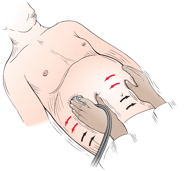
"Succusion splash” technique for assessing distention of abdominal
viscera
.

Cause

Pathophysiology of ascites: increased hepatic resistance to portal flow leads to portal hypertension. The splanchnic vessels respond by increased secretion of nitric oxide causing splanchnic artery vasodilation. Early in the disease increased plasma volume and increased cardiac output compensate for this vasodilation. However, as disease progresses the effective arterial blood volume decreases causing sodium and fl uid retention through activation of the renin-angiotensin system. The change in capillary pressure causes increased permeability and retention of fluid in the abdomen.


Differential Diagnosis

  • Chronic parenchymal liver disease, leading to portal hypertension
  • Peritoneal carcinomatosis
  • Congestive heart failure
  • Peritoneal tuberculosis
  • Nephrotic syndrome
  • Pancreatitis
Laboratory Tests
  • Initial evaluation should always include:
    • Diagnostic paracentesis. Laboratory tests on this fluid should include a CBC with differential, albumin, total protein, culture and a Gram stain. Optional tests on paracentesis fluid (depending on patient’s history) include amylase, LDH, acid-fast bacilli and glucose levels
    • AST, ALT, total and direct bilirubin, albumin, alkaline phosphatase, GGTP
    • CBC, coagulation studies
    • Electrolytes, BUN, creatinine
  • A serum to ascites albumin gradient (SAAG) should be calculated in all patients. If the SAAG is greater than 1.1, the cause of ascites can be attributed to portal hypertension. If SAAG is less than 1.1, a nonportal hypertension etiology of ascites must be sought.
Imaging studies
  • Endoscopy of the upper GI tract to evaluate for esophageal varices if ascites is secondary to portal hypertension.
  • Abdominal ultrasound is the most sensitive measure for detecting ascitic fl uid; a CT scan is a viable alternative.
  • Liver biopsy in select patients (i.e., those with portal hypertension of uncertain etiology).
Treatment
  • Sodium-restricted diet (maximum 60-90 milliequivalents per day).
  • Fluid restriction to 1 liter per day in patients with hyponatremia.
  • Patients with moderate-volume ascites causing only moderate discomfort may be treated on an outpatient basis with the following diuretic regimen: spironolactone 50-200 mg daily or amiloride 5-10 mg daily. Add furosemide 20-40 mg per day in the fi rst several days of treatment, monitoring renal functions carefully for signs of prerenal azotemia (in patients without edema goal weight loss is 300-500 grams/ day, in patients with edema 800-1000 grams/day).
  • Patients with large-volume ascites causing marked discomfort or decrease in activities of daily living may also be treated as outpatients if there are no complications. There are two options for treatment in these patients: (1) largevolume paracentesis or (2) diuretic therapy until loss of fluid is noted (maximum spironolactone 400 mg daily and Lasix 160 mg daily). There is generally no difference in longterm mortality; however paracentesis is faster, more effective,
    and associated with fewer adverse effects.
  • Five percent to 10% of patients with large-volume ascites will be refractory to high-dose diuretic treatment. Treatment strategies include repeated large-volume paracentesis with infusion of albumin every 2-4 weeks or placement of a transjugular intrahepatic portosystemic shunt (TIPS).
Read More

Proteins


Proteins are created from linear polymers of amino acids linked to each other via peptide bonds.
  • Oligopeptides: small peptide polymers containing between 2 and 50 amino acids
  • Polypeptides: large peptide polymers containing more than 50 amino acids
Although 20 amino acids are used to construct proteins, 10 of them are considered essential (including arginine, which is essential in growing children), because they cannot be synthesized de novo and therefore must be obtained from the diet. They can be remembered with the mnemonic PVT TIM HALL, which refers to 
Phenylalanine             Threonine           Histidine
Valine                           Isoleucine           Arginine
Tryptophan                 Methionine         Leucine
                                                                     Lysine


Proteins from animal sources contain all 10 essential amino acids and are therefore considered high quality. Proteins from plant sources, however, contain some but not all essential amino acids. A judicious combination of different plant sources can provide a balanced supply of high-quality protein.

Digestion and Absorption

The acidic pH (< 2) of the stomach denatures proteins and thus makes their polypeptide chains more susceptible to enzymatic degradation by peptidases, as follows:
  • Pepsin: Proteins are initially digested in the stomach by the gastric enzyme pepsin. It is most effective at low pH and is inactive at a higher pH such as that observed in the duodenum (pH > 7).
  • Pancreatic peptidases: The short polypeptides generated from the action of pepsin are further cleaved in the duodenum by pancreatic peptidases. Cleavage by these peptidases releases amino acids, which are absorbed by intestinal cells.
Function

Proteins are a major source of fuel, enzymatic activity, and structural support (e.g., keratin, collagen). Their degradation into amino acids provides the building blocks for the synthesis of other important molecules (e.g., heme molecules and nucleotides).       
Read More

Lipids

Lipids

Ingested lipids consist of a heterogeneous mixture of hydrophobic particles that includes the following: 
  • Fatty acids: These monocarboxylic acids, which contain hydrocarbon chains, are either saturated (zero double bonds), monounsaturated (one double bond), or polyunsaturated (two or more double bonds).
  • Triacylglycerols: These lipids are composed of three fatty acids attached to a glycerol backbone. They are stored in adipose tissue as fat.
  • Cholesterol and cholesterol esters: These hydrophobic molecules respectively consist of free cholesterol and a cholesterol molecule esterified to a fatty acid.
  • Membrane lipids: Phospholipids are found in cellular membranes and are composed of fatty acids attached to a glycerol backbone (glycerophospholipids) or a sphingosine backbone (sphingophospholipids). A phosphate group is esterified to the glycerol and sphingosine backbones. Other membrane lipids, called glycolipids, include those that contain a fatty acid chain attached to a sphingosine backbone, which is in turn attached to carbohydrate residues.
  • Lipid-soluble vitamins: These include vitamins A, D, E, and K.

Lipids Structure
Digestion and Absorption
 
The digestion of lipids is catalyzed by lipases (enzymes that hydrolyze ester bonds) and is aided by the lipid-emulsifying molecules in bile. 
  • Salivary (lingual) lipase and gastric lipases break down only short-chain and medium chain fatty acids. Short-chain fatty acids then diffuse through the stomach to enter the circulatory system.
  • Bile acids/salts are amphipathic molecules that emulsify dietary lipids in the duodenum, making it easier for digestive enzymes (released by the pancreas and cells lining the intestines) to hydrolyze them. Hydrolysis of ester bonds in triacylglycerols, cholesterol esters, and phospholipids are catalyzed by lipases, cholesterol esterases, and phospholipases, respectively.
The lipid-soluble vitamins, as well as the products of lipid digestion—free fatty acids, 2-monoacylglycerols, and cholesterol— are assembled into spherical particles called micelles. Monomeric lipids are absorbed from the micelles by the villi of the intestinal mucosa. In these cells, the fatty acids are reassembled into triacylglycerols phospholipids, and cholesterol esters. The reassembled lipids are then incorporated into large spherical lipoproteins called chylomicrons for delivery to the liver.   
Lipid Digestion
Function
 
Lipids are important long-term energy providers, yielding 9 kcal/g. They are also the major structural components of cell membranes (e.g., phospholipids, glycolipids, and cholesterol). Lipids are used to generate lipid-soluble molecules, such as steroid hormones, bile acids, and eicosanoids, and the lipid-soluble vitamins A, D, E, and K. Several enzymes require lipids for their activity. 
Read More

Carbohydrates


Carbohydrates are the most plentiful energy source in the human diet and are available in the following forms:
  • Monosaccharides: These simple sugars, such as glucose and fructose, are found in fruits and honey.
  • Disaccharides: These carbohydrates, such as sucrose, lactose, and maltose, are composed of two monosaccharide units linked via a glycosidic bond. Sucrose is found in table sugar and fruits, lactose is found in milk, and maltose is found in malt sugar.
  • Oligosaccharides: These carbohydrates are composed of from 3 to 10 monosaccharide units and are found in vegetables. Oligosaccharides are also found attached to proteins and lipids (e.g., glycoproteins and glycolipids, respectively) on the outer surface of cellular membranes in animals.
  • Polysaccharides: These carbohydrates are composed of more than 10 monosaccharide units and are exemplified by glycogen (from animal products) and starch (from plant products).
Classification of Carbohydrates
Digestion and Absorption

Various enzymes that contribute to the digestion and absorption of carbohydrates as they pass from the mouth to the intestines are as follows:
  • α-Amylase: The breakdown of polysaccharides into oligosaccharides and disaccharides is catalyzed by salivary α-amylase (in the oral cavity) and pancreatic α-amylase (in the duodenum).
  • Lactase, sucrase, maltase: Oligosaccharides and disaccharides are hydrolyzed into monosaccharides by the enzymes lactase (which targets lactose), sucrase (which targets sucrose), and maltase (which targets maltose). These enzymes are produced and secreted by intestinal cells. The resulting monosaccharides (e.g., glucose, galactose, and fructose) are absorbed by epithelial cells of the intestinal mucosa and transported by the portal circulation to target tissues.
Functions
Glucose, the most important molecule for energy, is stored in the liver and muscle as glycogen, which can be degraded when energy is needed. Carbohydrates are also important components of cellular membranes (as components of glycolipids and glycoproteins), tissues (as components of bones and cartilage), and genetic material (as the sugar molecules in nucleic acids). 
 
Read More